

What physicians should know about recognizing attacks, finding secondary disease, and knowing when to refer to rheumatology.
Raynaud's phenomenon is easy to dismiss as simply having cold hands.
Most cases are benign. But in some patients, these color changes are an early clue to an underlying connective tissue disease or a vascular problem.
The goal of this article is to help you recognize a Raynaud's attack, identify the pattern that is likely benign, and understand what should trigger a closer look.
1. Start With the Attack
Raynaud's is an episodic vasospasm, usually triggered by cold exposure or emotional stress.
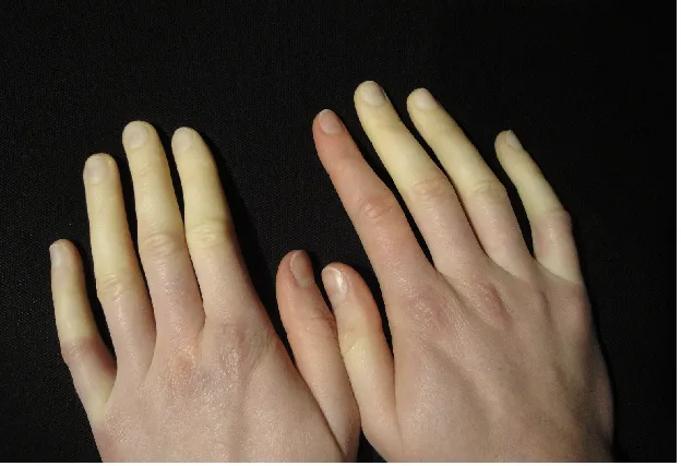
Patients describe sharply demarcated pallor or discoloration in one or more digits, followed by numbness, tingling, throbbing, or redness as blood flow returns. It is important to note that Raynaud's can affect both the fingers and the toes.
A common misconception is that the fingers must change to all three colors: white, then blue, then red. They do not. A single color change is enough to raise suspicion for Raynaud's.
In darker skin tones, the change may be subtler, so compare digits and ask about temperature changes, numbness, and pain.
Because the hands may look normal by the time the patient reaches clinic, ask the patient to photograph an attack. A well documented picture is often the most important diagnostic clue.
2. Identify the Pattern That Looks Primary
Primary Raynaud's is the more common form and is generally benign.
The reassuring patterns for Raynaud's are: young onset, symmetric attacks, complete resolution with rewarming, normal peripheral pulses, and a normal examination between episodes.
Primary Raynaud's does not cause fingertip ulcers, pitted scars, gangrene, or tissue loss.
Not every patient with the primary pattern needs an extensive autoimmune panel or an automatic rheumatology referral.
Reassurance and routine monitoring with primary care is usually appropriate for primary Raynaud's.
3. Know What Does Not Fit
Secondary Raynaud's, on the other hand, occurs because of another disease, medication, exposure, or vascular process. Connective tissue diseases, especially systemic sclerosis, but also lupus, Sjögren's disease, inflammatory myositis, and rheumatoid arthritis, are important causes.
The pattern deserves further evaluation when: symptoms begin after age 30, become severe or progressive, affect one hand or a single digit, involve the thumb, or fail to resolve completely.
Red flags that warrant further investigation: Digital ulcers, pits, delayed healing, abnormal pulses, or persistent pallor or cyanosis.
In patients with suspected secondary Raynaud's, look beyond the color change. Examine for puffy fingers, skin thickening, telangiectasias, abnormal nailfold capillaries, inflammatory arthritis, rashes, or muscle weakness. Ask about reflux or dysphagia, dyspnea, sicca symptoms, photosensitivity, oral ulcers, and constitutional symptoms.
Also review the medication list closely: stimulants, some beta-blockers, vasoconstrictive migraine therapies can be possible culprits. A social history is crucial too, as nicotine use and exposure to vibrating tools worsen Raynaud's.
4. Order Tests to Answer a Question
For a patient with Raynaud's, labs are most helpful when assessing for secondary causes.
If the history or examination suggest an underlying cause, a reasonable evaluation includes complete blood count, complete metabolic panel, urinalysis, ESR, CRP, TSH, and ANA with reflex testing.
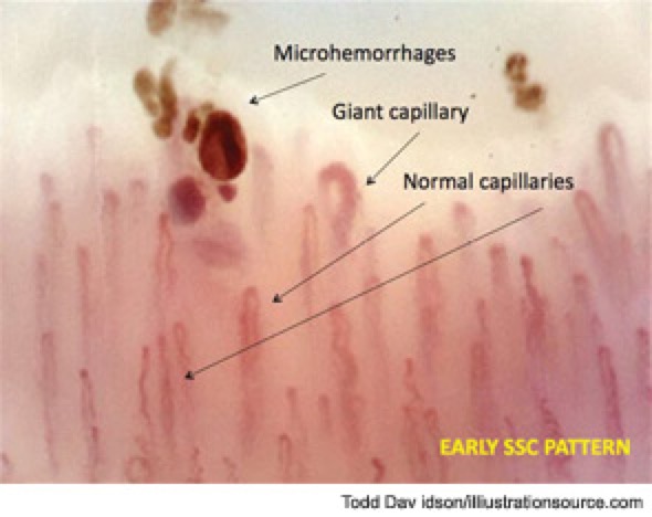
Nailfold capillaroscopy, usually performed by a rheumatologist, can be especially useful when systemic sclerosis or another connective tissue disease is suspected. Dilated or distorted capillary loops, hemorrhages, and capillary dropout support the presence of secondary Raynaud's.
5. Treat the Symptoms
For uncomplicated primary Raynaud's, begin with measures that prevent vasospasm: keep the whole body warm, use gloves when handling refrigerated items, avoid abrupt temperature changes, stop nicotine, and review vasoconstrictive medications.
When medication is needed for Raynaud's, set expectations. The goal is fewer, shorter, and less painful attacks, not necessarily the complete elimination of color changes.
Long acting dihydropyridine calcium channel blockers are first line. Nifedipine has the strongest evidence, but amlodipine is commonly used and may be easier to tolerate. Start with a low dose and titrate gradually toward the maximum tolerated dose. Hypotension, headache, flushing, and peripheral edema often limit treatment.
If symptoms do not improve after an adequate trial of calcium channel blockers, first confirm adherence, trigger control, and whether the presentation still appears primary before adding another vasodilator.
Topical nitroglycerin may help when one or two digits are particularly symptomatic, although headache and hypotension can limit its use.
For refractory Raynaud's, phosphodiesterase-5 inhibitors such as sildenafil may be considered, generally in collaboration with rheumatology.
6. Decide Who Needs Reassurance, Further Evaluation, or Urgent Care
When a patient presents with suspected Raynaud's, use four questions to determine what should happen next:
Confirm the attack: Is the color change episodic, triggered by cold or stress, and sharply demarcated?
Map the pattern: Is it symmetric and fully reversible?
Look for ischemic injury: Are there digital pits, ulcers, delayed healing, severe pain, or fixed discoloration?
Check the context: Are peripheral pulses normal? Are there systemic symptoms, abnormal nailfold findings, vasoconstrictive medications, nicotine use, or occupational exposures?
Then place the patient into one of three pathways:
- Likely primary Raynaud's: Symmetric, fully reversible attacks with normal pulses, no tissue injury, and no concerning systemic findings. Begin conservative management and monitor for change.
- Possible secondary Raynaud's: Atypical, asymmetric, severe, progressive, or later-onset symptoms warrant further testing and rheumatology referral.
- Possible digital ischemia: Persistent pallor or cyanosis, severe pain, ulceration, infection, or tissue loss requires urgent assessment by a vascular surgeon.
The clinical task is not just to confirm Raynaud's.
It is to determine whether the vasospasm is benign, disease-associated, or threatening the digit, then treat accordingly.
Sources
- Niklas D. Raynaud-Syndrom.JPG. Wikimedia Commons. CC BY-SA 3.0.
- https://www.the-rheumatologist.org/article/capillaroscopy-a-safe-and-direct-method-for-ssc-diagnosis/
- National Institute of Arthritis and Musculoskeletal and Skin Diseases. Raynaud's Phenomenon: Diagnosis, Treatment, and Steps to Take.
- American College of Rheumatology. All About Raynaud's Phenomenon.
- Herrick AL. Raynaud's phenomenon. Journal of Scleroderma and Related Disorders.
- Smith V, et al. Standardisation of nailfold capillaroscopy for the assessment of patients with Raynaud's phenomenon and systemic sclerosis. Autoimmunity Reviews. 2020;19(3).
- Shapiro SC, Wigley FM. Treating Raynaud phenomenon: Beyond staying warm. Cleveland Clinic Journal of Medicine. 2017;84(10):797–804.